The structure of DNA: the double helix
“In 1953 Watson and Crick showed that DNA is a double helix, two chains joined by paired bases, using X-ray data from Rosalind Franklin and Maurice Wilkins; the pairing showed how genes are copied.”
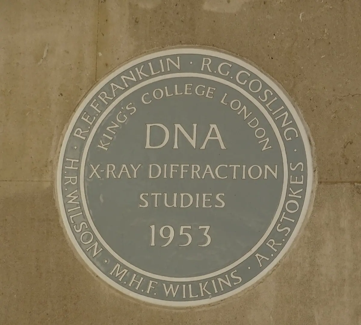
In 1953 two young scientists at the Cavendish Laboratory in Cambridge, the 24-year-old American biologist James Watson and the 36-year-old British physicist Francis Crick, worked out the shape of DNA, the molecule that genes are made of. It is a double helix: two long chains of sugar and phosphate wound round each other, running in opposite directions and held together in the middle by pairs of bases, adenine always with thymine and guanine always with cytosine. Their short paper in Nature of 25 April 1953 was printed beside two papers from King's College London that gave the X-ray evidence, one by Maurice Wilkins and two colleagues, the other by Rosalind Franklin and her student Raymond Gosling.
The structure explained at once how life copies itself. Because each base has only one partner, the order of bases along one chain fixes the order along the other; if the chains come apart, each can serve as the pattern for a new partner, and two identical molecules result. Watson and Crick hinted at this in a famous understatement, that the pairing 'immediately suggests a possible copying mechanism for the genetic material', and spelled it out in a second paper on 30 May. The order of the bases, they saw, could be a code, and within a decade Crick and others showed that it spells out proteins three bases at a time.
The way there was a race. Since Oswald Avery's experiments of the 1940s there had been good reason to think genes were made of DNA, but nobody knew its shape. At King's, Wilkins and, from 1951, Franklin photographed DNA fibres with X-rays; Franklin sorted out its two forms, a drier 'A' and a wetter 'B', and in May 1952 she and Gosling took the sharpest picture yet of the B form, 'Photograph 51', whose cross of spots is the mark of a helix. In Cambridge, Watson and Crick tried to guess the structure by building models, as the American chemist Linus Pauling had done for proteins. Their first try, in late 1951, put the backbones in the middle, and Franklin showed at once that it was wrong. Early in 1953 Pauling sent out a three-chain structure with the same flaw. Spurred on, Watson went to King's, where Wilkins showed him Photograph 51, and soon afterwards the pair were handed a report with Franklin's measurements. By the end of February Watson, playing with cardboard cut-outs, had seen how the bases pair, in early March they had their metal model, and in April Pauling himself agreed that they were right.
Who deserves the credit has been argued ever since. Franklin did not know that her photograph had been shown to Watson, and her data reached Cambridge in a report that was not confidential; her own draft of 17 March 1953 already called a helix 'highly probable'. She moved to Birkbeck College, did important work on the structure of viruses, and died of cancer in 1958, aged 37. In 1962 Watson, Crick and Wilkins shared the Nobel Prize in Physiology or Medicine. Watson's lively but one-sided memoir The Double Helix (1968) made Franklin a caricature, and later writers turned her into the story's wronged heroine; historians who have read her notebooks now see her as an equal member of the group of four whose work solved the structure.
The double helix became the foundation of molecular biology: the genetic code, the reading of whole genomes and the editing of genes all rest on it. Odile Crick's simple drawing of it in the 1953 paper, two ribbons twisting round an axis, became one of the best-known images in science.
What happened
- 1951
Rosalind Franklin, a physical chemist trained in X-ray diffraction in Paris, joins John Randall's Medical Research Council biophysics unit at King's College London, where Maurice Wilkins is deputy. The two soon clash, and Randall divides the DNA work, Wilkins handing her the unit's purest DNA.
King's College London, the Strand: the biophysics unit of John Randall, Maurice Wilkins, Rosalind Franklin and Raymond Gosling, where Photograph 51 was made in May 1952At a symposium at the Zoological Station in Naples late in May, the young American biologist James Watson meets Maurice Wilkins and sees for the first time an X-ray pattern of DNA, a sign that the molecule has a regular structure. He resolves to work on it.
Stazione Zoologica Anton Dohrn, Naples: where Watson first saw an X-ray pattern of DNA, shown by Wilkins at a symposium in May 1951Watson starts work at the Cavendish Laboratory in Cambridge, in the Medical Research Council unit led by Max Perutz, and shares a room with Francis Crick, a physicist twelve years older who is still a research student. They agree to find DNA's structure by building models, as Linus Pauling had found the alpha helix of proteins.
The Cavendish Laboratory, Free School Lane, Cambridge: home of the Medical Research Council unit where Watson and Crick shared a room and built their modelsFranklin gives a colloquium at King's on her X-ray work, which Watson attends. From his memory of it he and Crick build a three-chain model with the phosphate backbones in the middle. Invited to Cambridge to see it, Franklin points out that DNA holds far more water than the model allows and that the phosphates must lie on the outside. Lawrence Bragg, head of the Cavendish, tells the pair to drop DNA.
The Cavendish Laboratory, Free School Lane, Cambridge: home of the Medical Research Council unit where Watson and Crick shared a room and built their models- 1952
Franklin and Gosling set up their camera on Friday 2 May and develop the film on Tuesday 6 May, after 62 hours of exposure: 'Photograph 51', number 51 in a series and the clearest picture yet of the wet B form of DNA, with the cross-shaped pattern of a helix. Franklin puts it aside to concentrate on the drier A form.
King's College London, the Strand: the biophysics unit of John Randall, Maurice Wilkins, Rosalind Franklin and Raymond Gosling, where Photograph 51 was made in May 1952Unable to fit the crystalline A form to a helix, Franklin and Gosling send Wilkins a mock funeral card announcing the 'death' on Friday 18 July 1952 of the crystalline DNA helix.
King's College London, the Strand: the biophysics unit of John Randall, Maurice Wilkins, Rosalind Franklin and Raymond Gosling, where Photograph 51 was made in May 1952At Caltech in Pasadena, Linus Pauling and Robert Corey send off their paper 'A proposed structure for the nucleic acids': three chains wound together with the phosphates in the middle. It appears in February 1953 in the Proceedings of the National Academy of Sciences.
California Institute of Technology, Pasadena: Linus Pauling's laboratory, where he and Robert Corey proposed a three-chain structure in December 1952- 1953
A copy of Pauling's manuscript reaches Cambridge through his son Peter. Watson and Crick spot a basic chemical slip: his phosphates keep hydrogen atoms that DNA, an acid, loses, and without them his three chains would fly apart. Watson takes the news to King's, where Wilkins, who has a print from Gosling now that Franklin is about to leave for Birkbeck College, shows him Photograph 51.
King's College London, the Strand: the biophysics unit of John Randall, Maurice Wilkins, Rosalind Franklin and Raymond Gosling, where Photograph 51 was made in May 1952Bragg lets Watson and Crick return to DNA. Max Perutz passes them a report on the King's unit written for a Medical Research Council visit in December 1952; Franklin's page confirms a repeat every 34 ångströms and gives the symmetry of her crystals, which, Crick sees, means chains running in opposite directions.
The Cavendish Laboratory, Free School Lane, Cambridge: home of the Medical Research Council unit where Watson and Crick shared a room and built their modelsMoving cardboard cut-outs of the bases, after the American chemist Jerry Donohue has corrected their shapes, Watson sees that adenine pairs with thymine and guanine with cytosine, which explains Erwin Chargaff's ratios. On Saturday 28 February, as Cambridge tells it, Crick and Watson celebrate at the Eagle, Crick announcing that they have found the secret of life.
The Eagle, Bene't Street, Cambridge: the pub near the laboratory where Watson and Crick celebrated on 28 February 1953In early March Watson and Crick build their model in metal: two right-handed chains coiled round one axis, the phosphates outside and the paired bases stacked inside, 3.4 ångströms apart and ten to a turn.
The Cavendish Laboratory, Free School Lane, Cambridge: home of the Medical Research Council unit where Watson and Crick shared a room and built their modelsAt King's, unaware of the Cambridge model, Franklin and Gosling finish a rough draft on the B form: a helical structure, most likely two chains with the bases inside and the phosphates outside, is 'highly probable'.
King's College London, the Strand: the biophysics unit of John Randall, Maurice Wilkins, Rosalind Franklin and Raymond Gosling, where Photograph 51 was made in May 1952Crick writes a seven-page letter to his twelve-year-old son Michael at boarding school, telling him that he and Watson have probably made a most important discovery, with a sketch of the helix and the 'copying mechanism by which life comes from life'.
Watson and Crick send Pauling their typescript with a letter saying they could not suppress their doubts about his structure. In the same weeks Wilkins, who has seen their paper, asks that the King's results appear in the same issue of Nature, his own group's and Franklin and Gosling's.
Pauling, in England on his way to the Solvay conference in Brussels, sees the model and Franklin's photograph in Cambridge and writes to his wife on 6 April that his structure is probably wrong and theirs right. At the conference Bragg gives the first public announcement of the double helix.
The Cavendish Laboratory, Free School Lane, Cambridge: home of the Medical Research Council unit where Watson and Crick shared a room and built their modelsNature prints the three papers one after another: Watson and Crick's structure, with Odile Crick's drawing; the X-ray evidence of Wilkins, Stokes and Wilson; and Franklin and Gosling's, with Photograph 51.
About three weeks later Bragg lectures on the discovery at Guy's Hospital Medical School in London, and the News Chronicle puts it on its front page. A draft article for Time, written with Franklin's help, describes two teams working together, but is never printed.
Watson and Crick's second paper, 'Genetical implications of the structure of deoxyribonucleic acid', explains how the two chains could separate, each serving as the pattern for a new partner, so that a gene is copied exactly.
At the Royal Society's summer conversazione in London, one of the first public displays of the double helix is signed by the authors of all three Nature papers, with Franklin as lead author: at the time the discovery is seen as the work of the King's and Cambridge groups together.